Super-resolution Imaging
Stimulated Emission Depletion Microscopy
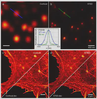
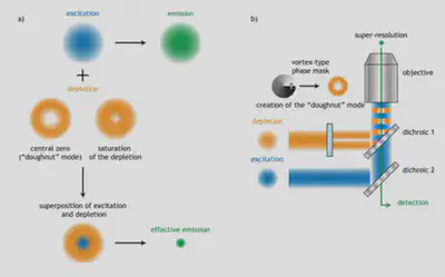
An intuitive understanding of STED microscopy (see Figure 1) can be realized as the action of two laser beams on the dynamics of a fluorescent molecule (fluorophore) that is attached to the subcellular structure. The two laser beams are spectrally se-lected to match the absorption spectrum (Excitation beam) and the red edge of the emission spectrum (STED beam) of the fluorophore. Conventionally, the excitation beam is focused down to a spot and induces an excitation and a subsequent relaxa-tion of the fluorophore molecule by the spontaneous emission of a fluorescent pho-ton. This spot is scanned across the sample to build an image of the sample. Hence the resolution is governed by the size of this diffraction limited focal spot. To over-come this limit, the STED beam is shaped in the shape of a doughnut and its focal spot is overlapped spatially with the excitation volume. The role of the STED beam is to control spatially and temporally the rate of depletion of this excited state before any fluorophore emission occurs. This is done by forcing all the molecules at the pe-riphery of the doughnut to ground state instantaneously by stimulated emission. Meanwhile those in the central dark spot continue to emit fluorescence. By simply increasing the number of incident STED photons, one can saturate this depletion of the excited state, thereby decreasing the effective detection volume to well below the diffraction limit
The key challenges towards realizing and building systems were two-fold i) the interferometric precision involved in its operation, considering two or three ultrashort pulses with low temporal coherence and ii) the variability involved in working with complex biological and chemical systems. I was involved in practically all aspects of the project and set up such a system from scratch. The guiding design principles were robustness and user-friendly behavior since the system was to be hosted at an imaging platform (Centre de Photonique Biomédicale, Orsay). A pulsed super-continuum fiber laser was employed to ensure flexibility, low average powers compatible with bio-imaging and time resolved measurements. Different strategies with respect to shaping the depletion beams such as the use of spatial light modulators and phase masks on the physical substrates were explored. A large part of my work was to identify and solve the bottlenecks of using commercially available black-box hardware. Through the course of first two years of the dissertation, these issues were addressed with a combination of home-built systems for scanning and acquisition. In addition, we also developed a time resolved detection system, based on Time Correlated Single Photon Counting, to examine changes to the lifetime of the fluoro-phores in different conditions. At this time, this was the first demonstration of such time-resolved STED systems at the national level.

Super-critical Angle Fluorescence Microscopy
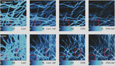
In addition to lateral resolution, a number of biological studies also require a high axial resolution. This has proved particularly challenging to address over the years. For instance, in membrane biology, this question reduces to the optical confinement of the light fields to a sub diffraction limited zone at the membrane interface. In a novel alternative approach to imaging the cellular membrane, we detect exclusively what is known as the supercritical angular fluorescence (SAF). When an emitter is sufficiently close to an interface between two media with different refractive indices, the near field components of radiation can tunnel through and become propagative on the side with the higher refractive index. This appears as light well above the critical angle as defined by Snell-Descartes law. This supercritical angular emission can be up to 34% of the total emission when the molecule is on the surface and exponentially decreases when it is further away. Hence, this allows us to selectively image the membrane by detecting purely the SAF signal.

Spot Variation STED-FCS
As one of the few experts on the hands-on setup of STED systems, I had the oppor-tunity to collaborate with group of Didier MARGUET (CIML, Marseille) on a novel correlation spectroscopy technique, called spot variation fluorescence correlation spectroscopy (svFCS). The strength of said technique is to allow the study of diffu-sion and motility on biological surfaces at varying spatial scales. A particular scien-tific goal was to determine if the so-called transient nano-domains are simply arti-facts since information was not available at multiple length scales (nano-metric to hundreds of micrometers). In this regards, we aimed to push the upper-bound on the resolution to tens of nanometers by combining svFCS to STED. This instrument, fusing both imaging and correlation spectroscopy at multiple length scales, is now hosted on the Luminy campus of the Aix-Marseille Université.